Learn about alignment and shift
The next thing you should do is learn about what’s called
‘shifting the beam’ and aligning an aperture. As an
experimental electron microscopist, you will spend many
happy hours of your life shifting beams and aligning
apertures.
Ask the demonstrator: To set up the microscope with no specimen,
condenser system aligned, but now put in and align a small
condenser aperture. Show me how to shift the beam.
You will be shown two knobs, which will probably be labelled
‘shift x’ and ‘shift y’. They are sometimes simply
referred to as the ‘electrical
shifts’. Focus C2 so that you can see a tiny in-focus image
of the filament as before.
Experiment: Now over-focus C2 (turn the brightness knob
clockwise) until there is a small circle on the phosphor
screen. Try turning the shift knobs. The circle moves
around the place. The two shift knobs refer to the two
Cartesian co-ordinates of the image plane, and they should
be at right-angles to each other. (The movements may not be
perfectly at right angles, for reasons which are too
complicated to explain at this stage).
Try changing the focus of C2 while the beam is shifted to
one edge of the phosphor screen. It should behave exactly
as before, but just shifted in position. Easy.
Ask the demonstrator: To show me how to move the condenser
aperture.
You will be shown two mechanical screw adjustments on a
block sticking out of the electron microscope. The thing is
called an ‘aperture mechanism’ or ‘aperture assembly’ and it
controls the position of a tiny hole (typically 10-500 microns in
diameter) which is supposed to sit on the centre-line of the
whole microscope.
The centre line of the entire microscope is called the
‘optic axis’, and it is essential that unless you are
deliberately ‘tilting the beam’ (which we will come to
later), all the lenses and apertures of the microscope are
centred on the optic axis.
There are three aperture mechanisms on the column. For the
time being, lets just understand the condenser aperture,
which is the one at the top of the column.
Before touching the condenser alignments, slightly over-
focus C2, as before, and use the shifts to get the disc into
the very centre of the phosphor screen.
Now try moving the condenser aperture: the disc shifts. In
fact the disc is the shadow cast by the condenser aperture.
Shift the disc by moving the aperture right to one side of
the phosphor screen.
Use the ‘shift’ knobs - the electrical knobs you have just
learnt about - to move the disc back to the centre of the
screen.
Now change C2. What’s happening? The disc moves and comes
in and out of focus. You’ll find that you can easily ‘lose
the beam’ - that is, get into a situation where you can’t
see anything on the phosphor screen at all, no matter how
much you change the position of the aperture or the beam
shifts. This is good way of annoying the demonstrator, if you
are in that sort of mood...
Ask the demonstrator: To get the beam back as it was before.
You should now have a few moments to think about what’s
going on.
Theory: The useful part of an electron lens is extremely
small – only the centre tens of microns of an electron lens
is any good at focussing electrons. For this reason, we
have to use apertures. We can think of the condenser
aperture sitting immediately below the lens we have been
playing with, C2.
When the demonstrator set up the microscope, this is what the ray
diagram looked like:
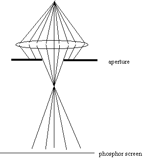
Beams come out of the filament in a huge range of angles,
but only a few of these get through the condenser aperture
and onto the phosphor screen. When you moved the aperture
to one side, this is what happened:
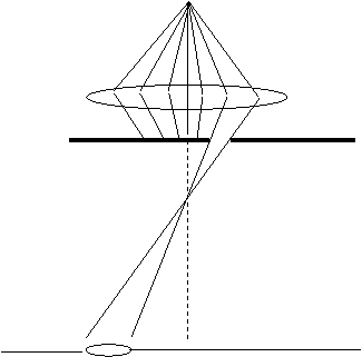
Now what happens when you change the focus of C2? Well, the
focal point of the lens moves up and down vertically, as
before. But because the aperture is so offset to one side,
the place where the beam hits the phosphor moves laterally,
as well as going in and out of focus. It seems complicated
when you see it happen: in fact its very simple when you
think of the ray diagram.
Experiment: Play around with the condenser aperture and C2
focus to see if you can understand the ray diagram in the figure
above.
Let us spend a little time thinking about the ‘shift x’ and
‘shift y’. You will use these knobs all the time when you
use the electron microscope. It is pretty important to
understand how they (and similar controls you will encounter
later) work.
Because the useful part of an electron lens is so tiny, it
is essential that each lens is aligned with the optic axis
(the very centre line of the microscope), preferable within
a few microns. It is virtually impossible to do this
mechanically (like we moved the condenser aperture), because
electron lenses are so heavy and unwieldy.
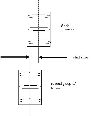
Supposing we have two halves of an electron microscope.
Both halves contain a number of lenses, all of which are
neatly lined up with another inside each half.
Unfortunately, the two halves are misaligned with respect to
each other, like this
It is easy to bend an electron beam by applying an electric
or magnetic field across the beam. Electric fields are
sometimes used in electron microscopes to deflect beams, but
usually a pair of magnetic coils are used, mounted opposite
one another. The same method is used in a television to
scan the electron beam over the surface of the screen. Such
coils are called ‘deflection’ or ‘alignment’ coils, and we
can draw them in a ray diagram by a pair of curly lines
either side of the beam. We could try to counteract the
misalignment shown above by using a pair coils thus
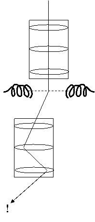
But we will quickly see that although the beam now passes
from the first half of the electron microscope column into
the second half, it now goes into the second half at a
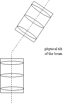
rather extreme angle. This is called a ‘tilt’. It has the
same effect as if the alignment coils did not exist, but the
column had been put together physically like this:
It turns out that sometimes we want to tilt the beam.
However, how do we solve the first problem of the physically
shifted column? The answer is to have two sets of coils,
like this:
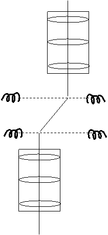
The first coils bend the beam to the left, the second bend
it back again by exactly the same amount. The net effect
corrects the original alignment fault. Later you will learn
that the we sometimes have to adjust the ratio of these
bending coils to make sure that they achieve exactly what we
want. (This adjustment is sometimes called changing the
‘rocking point’ of the deflection coils.) In a typical
transmission electron microscope there are about three or
more sets of such coils, which are usually called ‘double
deflection coils’. Of course, in reality each set of coils
have both x- and y-components, so that we can adjust the two
dimensions of possible misalignment.
The above diagram represents what we call a ‘pure shift’.
You should be warned that quite often knobs that are meant
to be ‘shift’ knobs also introduce of bit of ‘tilt’ - or
vice versa, depending on the exact geometry of the coils
relative to the lenses.
Back to the experiment:
Now that we know what an aperture is and how the shift coils
work, can we understand what was going on in the previous
experiment? Well, a particular combination of beam shift
and aperture shift could result in ray diagrams that look
like this:
We have a source, a lens, an aperture and a pair of
deflection coils between the aperture and the phosphor
screen which we can use to shift the beam.
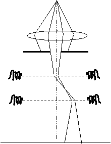
In the first diagram, the source, the lens and the aperture
are all aligned with respect to one another, but the place
where the beam hits the phosphor screen has been shifted by
the shift coils. If we change the focus of the lens, the
disc of the aperture will go in and out on its own axis, but
this axis is not the same as the centre of the phosphor
screen.
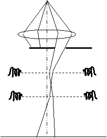
The only difference between the first and the second
diagram, is that in the second diagram the aperture has been
moved, so its circle appears to hit the phosphor screen in
the middle. But now both the aperture and the phosphor
screen (the electrical shifts) are out of line. The beam is
going through the side of the condenser lens.
It is very, very bad news when a beam goes through the side
of an electron lens; whenever possible, as many beams as
possible should pass beside the very centre of an
electron lens. This is because of another important
difference between optical lens and electron lenses;
electron lenses suffer from terrible ‘aberration’, which
seriously degrades any image we obtain, and which very
quickly gets very bad as the angle of a beam through a lens
increases.
Luckily, though, it is very easy to tell that an electron
lens is not lined up properly. As a general rule, if you
change the strength of any lens in the system and the image
(or diffraction pattern, whatever you’re looking at) moves,
then that lens is not lined up properly.
By now the demonstrator should have re-aligned the condenser
aperture.
Experiment: With the condenser aperture aligned, change C2.
See that the beam goes in and out of focus symmetrically:
the condenser is well-aligned - or at least it should be if
the demonstrator has done a good job. Now physically shift the
aperture (using the condenser aperture mechanism) say by
about its own diameter (the apparent diameter will depend on
the setting of C2). Shift the beam back to centre of the
phosphor screen using the beam shifts.
Looking at the circle on the phosphor screen like this,
could anyone tell whether or not the condenser aperture was
lined up? The answer is ‘no’ unless they are allowed to
alter C2.
Remember: If you want to test the alignment of a single
lens, alter its setting (i.e. its strength or excitation)
and see if anything moves.
Try it. Change C2 and you will see the centre of the shadow
of the aperture moves. That’s how you know that the
condenser aperture is not aligned.
Can you work out a way of re-aligning the condenser aperture
without asking the demonstrator? Look at the ray diagrams and
think about it. If you find a good way of doing it, then
you have learnt about half of everything you need to know
about electron beam alignment.
Most people learn to use electron microscopes by following
recipes or lists of commands or rules. You will never be a
truly good electron microscopist if you need a list of
commands. However, to get on quickly the next section, the
list of commands for aligning the condenser aperture is
this:
- Turn up the brightness knob until the illumination reaches the edge of the phosphor screen.
- Shift the condenser aperture to the centre of the phosphor screen using the mechanical shifts
on the condenser aperture assembly.
- Turn down the brightness knob until you see a focussed
spot, or until the illumination reaches the edge of the phosphor screen.
- Alter the electrical shifts to put the beam onto the centre of the phosphor screen.
- Repeat from (1) until the illumination appears to expand and contract around the centre of the phosphor screen.
Here’s another set of rules, which looks almost the same,
but doesn’t work at all:
- Turn up the brightness knob until the illumination reaches the edge of the phosphor screen.
- Alter the electrical shifts to put the beam onto the centre of the phosphor screen.
- Turn down the brightness knob until you see a focussed spot, or until the illumination reaches the edge of the phosphor screen.
- Shift the condenser aperture to the centre of the phosphor screen using the mechanical shifts on the condenser aperture assembly.
- Repeat from (1) until the illumination appears to expand and contract around the centre of the phosphor screen.
You would do well to draw the ray diagrams to work out why
one of these schemes works and the other doesn’t. But you
can do this at your leisure. In the meantime, the demonstrator
is anxious to get on with the lesson.
So far we have learnt about 5 variables: the focus of C2,
the electrical ‘x-shift’, the electrical ‘y-shift’, and two
controls on the condenser aperture assembly that control the
position of the aperture in two-dimensions.
In fact, you’ve now learnt nearly everything you need to
know about a single electron lens. The problem we tackle
next is how to put several electron lenses on top of one
another, and get them to work together.


Copyright J M Rodenburg
|